Skin cancer is the most common cancer in the UK, yet when caught early it is also one of the most treatable. As a consultant plastic surgeon with a specialist interest in skin oncology, I see first-hand how a timely assessment can transform the outcome for a patient.
This guide walks through the warning signs every adult should know, the three main types of skin cancer, and the practical situations in which a specialist review is worthwhile. Nothing here replaces a face-to-face examination, but it will help you decide when to pick up the phone.
The ABCDE rule: a simple framework for self-checks
The ABCDE rule is a widely used method for assessing pigmented lesions and moles. It is not a substitute for a dermatoscopic examination, but it is a genuinely useful tool for noticing changes early.
- A — Asymmetry: if you imagine drawing a line through the middle of the mole, the two halves should look broadly similar. Asymmetry is a soft warning sign.
- B — Border: benign moles usually have smooth, even borders. Ragged, notched, scalloped or blurred edges merit closer review.
- C — Colour: most reassuring moles are a single shade of brown. Multiple colours within one lesion — tan, dark brown, black, red, white or blue — should prompt attention.
- D — Diameter: lesions larger than 6 mm (the size of a pencil eraser) are more likely to warrant review, although melanomas can certainly be smaller.
- E — Evolving: the single most important letter. Any mole that is changing in size, shape, colour, texture or symptom (itching, bleeding, crusting) should be examined.
"If I could give one piece of advice, it would be this: trust change. A mole that was stable for years and is now doing something new is the single strongest reason to book a review."
The three main types of skin cancer
Most skin cancers fall into three categories, and each has a distinctive pattern of appearance.
Basal cell carcinoma (BCC)
BCC is the most common skin cancer and usually develops on sun-exposed areas such as the face, scalp, ears and back of the hands. It often appears as a pearly or waxy bump, a flat flesh-coloured or pale pink patch, or a small sore that scabs over, heals, and then breaks down again. BCCs tend to grow slowly and rarely spread, but they can be locally destructive if left untreated — particularly around the nose, eyes and ears.
Squamous cell carcinoma (SCC)
SCC typically presents as a firm, scaly or crusted patch, a persistent red bump, or a non-healing ulcer. It is more commonly seen in older adults with significant sun exposure, and on sites such as the lower lip, ears and backs of the hands. SCC can spread to lymph nodes if neglected, so prompt assessment of any persistent scaly lesion or non-healing sore is important.
Melanoma
Melanoma is less common than BCC and SCC but is the most serious form of skin cancer. It may arise within an existing mole or appear as a new pigmented lesion. The ABCDE features above are particularly relevant here. Melanomas can also develop in unexpected places, including the soles of the feet, under the fingernails, and in areas not regularly exposed to the sun, so a thorough head-to-toe check is valuable.
When to seek a specialist review
You should consider booking a skin assessment if any of the following apply:
- A changing lesion: any mole or patch that looks different from how it used to.
- A new lesion after the age of 40: most new pigmented lesions in middle age deserve a professional look.
- A non-healing sore: any area that fails to heal within four to six weeks.
- Persistent itching, bleeding or crusting: a mole that itches, oozes or bleeds without an obvious cause.
- A lesion that looks different from your others: the so-called "ugly duckling" sign — one mole that stands out from the rest of your pattern.
- A family history of melanoma: especially if you also have fair skin, many moles, or previous significant sun exposure.
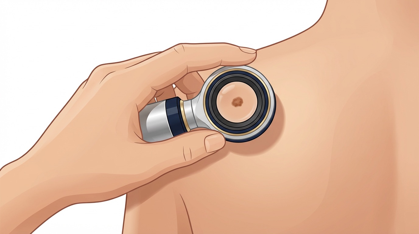
What to expect at a skin assessment
A specialist skin review typically combines a careful history, a full skin examination, and dermoscopy — a magnified, polarised view of the lesion that reveals structures invisible to the naked eye. If there is any doubt, a small excision biopsy under local anaesthetic will give a definitive answer. Most suspicious lesions can be fully removed in a single short procedure in an outpatient setting.
A word of reassurance
The vast majority of moles and skin changes are entirely benign, and many patients who come for review leave with simple reassurance. When something is found, early intervention usually means a small, straightforward procedure and an excellent prognosis. If you are unsure about a lesion, or if a mole is doing something new, please do not wait — a thirty-minute appointment is often all it takes to put your mind at rest.