Squamous cell carcinoma (SCC) is the second most common type of skin cancer in the UK, after basal cell carcinoma. It is diagnosed in tens of thousands of patients each year, and while most cases are entirely curable with straightforward surgery, SCC deserves a little more respect than its slower-growing cousin. Unlike basal cell carcinoma, squamous cell carcinoma can occasionally spread to the lymph nodes or beyond — which is why timely diagnosis and appropriate follow-up matter.
This guide explains what SCC is, how to spot it, who is most at risk, and what modern treatment involves.
What is squamous cell carcinoma?
SCC arises from the keratinocytes — the flat, plate-like cells that form most of the epidermis. When chronic ultraviolet damage or other triggers accumulate in these cells, they can lose their normal control over growth and begin to invade the tissues below. Once the cancer has broken through the basement membrane of the epidermis, it is classified as invasive SCC.
The vast majority of cutaneous SCCs are cured with local surgery alone. A minority — perhaps 4 to 5% — can spread, so recognising the higher-risk cases and treating them appropriately is the task of the specialist multidisciplinary team.
How SCC can appear
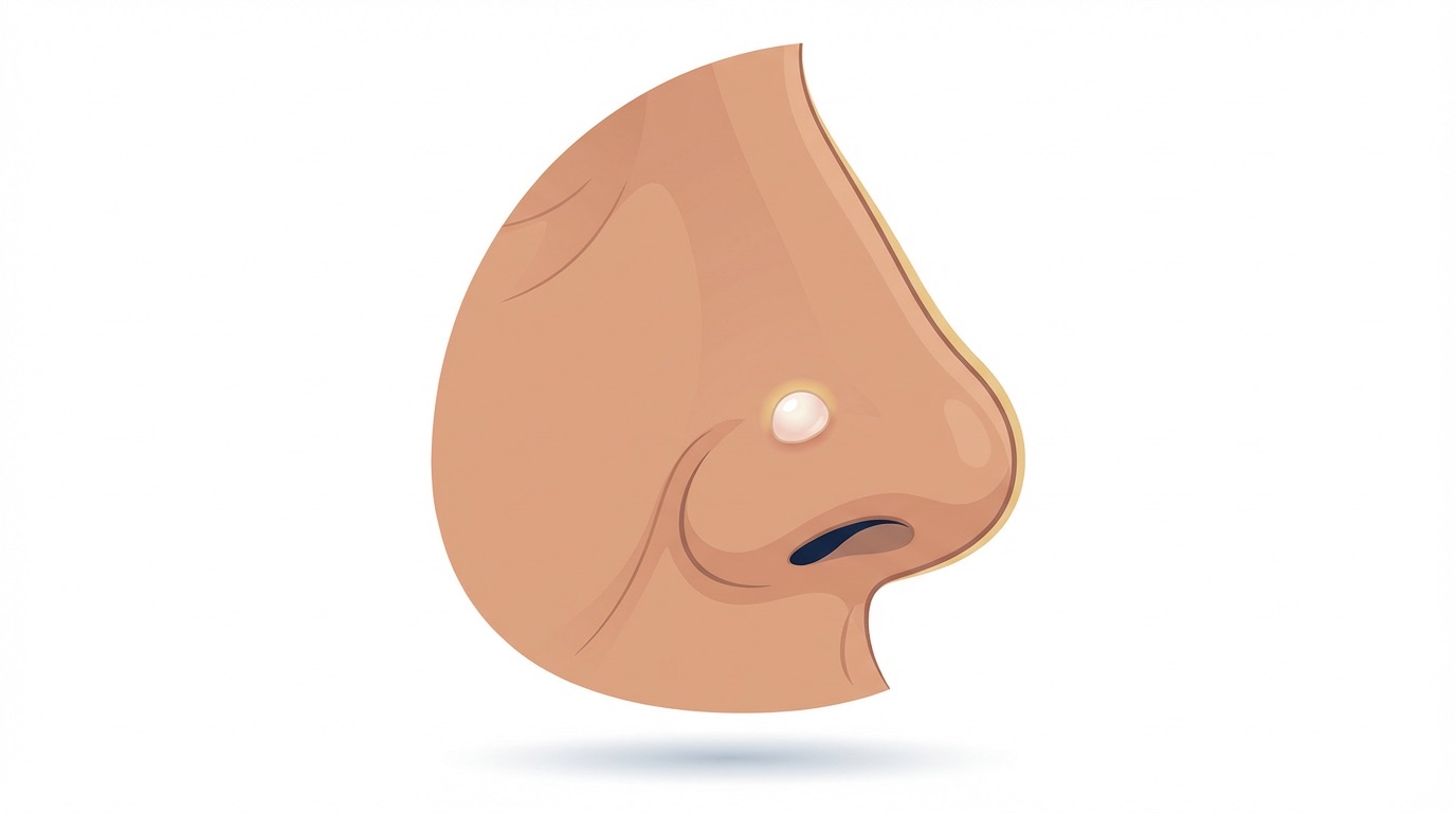
SCC rarely looks subtle. The classic clues include:
- A scaly, red patch that is rough to the touch and does not settle with moisturiser.
- A persistent, non-healing sore or ulcer — particularly one that bleeds on light trauma.
- A wart-like growth that is firm, tender, or grows surprisingly quickly over weeks.
- A crusted nodule with a horny central plug, sometimes raised above the surrounding skin.
Tenderness is a useful clue. Unlike benign lesions, SCCs are often surprisingly tender to palpation, and any sore spot that persists for more than a few weeks is worth a specialist look.
Precursor lesions
SCC often develops from recognisable pre-cancerous lesions. Treating these early can prevent invasive cancer altogether.
- Actinic keratoses: small rough, scaly patches on sun-exposed skin. Each individual lesion has a low risk of transformation, but patients with many are at higher risk overall.
- Bowen's disease: a form of SCC that is still confined to the epidermis (SCC in situ). It appears as a slowly enlarging, well-demarcated scaly pink patch — most often on the lower leg. Treatment at this stage is straightforward and prevents progression.
- Cutaneous horns: projecting columns of keratin that can overlie a benign lesion, actinic keratosis or SCC. Any cutaneous horn warrants biopsy of its base.
"A persistent scaly patch that keeps coming back despite moisturiser, or a sore that will not heal, deserves more than reassurance. A five-minute dermoscopic assessment can tell us whether to watch or to biopsy."
Who is at higher risk?
Several factors meaningfully increase the risk of SCC:
- Cumulative UV (Ultraviolet) exposure — the single biggest driver, including sunbed use.
- Fair skin that burns easily.
- Chronic wounds, burns and scars — SCC arising in an old burn scar or long-standing ulcer is known as a Marjolin's ulcer.
- Immunosuppression — organ transplant recipients have up to a hundredfold increased risk of SCC, and these tumours can behave more aggressively. Dedicated transplant skin surveillance clinics exist for precisely this reason.
- Human papillomavirus (HPV) infection in certain sites.
- Previous radiotherapy to the area.
- Occupational exposures such as arsenic, coal tar or long-term industrial UV.
High-risk sites
Certain anatomical locations carry a higher risk of behaving aggressively. These include the ears, the lips, the genital skin and any site of previous radiotherapy or chronic scarring. Tumours in these locations are more likely to be referred for specialist planning or Mohs surgery.
How SCC is diagnosed
Diagnosis begins with a careful history and skin examination, supplemented by dermoscopy. A confirmatory biopsy under local anaesthetic is almost always performed before definitive treatment so that the subtype, grade and depth of invasion can be assessed. For larger or higher-risk tumours, an ultrasound of the draining lymph nodes, or occasionally cross-sectional imaging, may be arranged.
Treatment options
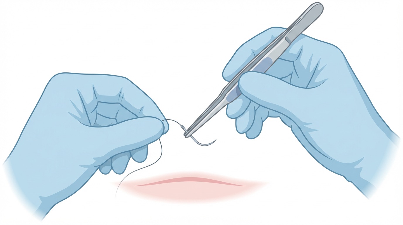
Wide local excision
The mainstay of treatment. The tumour is removed with a margin of normal skin — typically 4 to 10 mm depending on the risk category — under local anaesthetic as a day-case. The specimen is sent to the laboratory for histology, and the defect closed directly or reconstructed such as with a small flap or skin graft.
Mohs micrographic surgery
Reserved for high-risk SCCs: those on the central face, ears, lips, recurrent tumours or lesions in immunosuppressed patients. Mohs offers the highest cure rate while preserving as much healthy tissue as possible. Reconstruction is often performed on the same day — see our guide to Mohs reconstruction.
Radiotherapy
An option for patients unfit for surgery, for particular specialised anatomical sites such as the nose, or as an adjunct after surgery where margins are close or perineural invasion is present.
Adjuvant and systemic treatments
For advanced or metastatic SCC, immunotherapy (such as cemiplimab) has transformed outcomes over the last few years. Decisions about these treatments are made within a specialist skin cancer multidisciplinary team.
Follow-up after treatment
Follow-up is tailored to the risk category of your tumour, in line with the British Association of Dermatologists (BAD) guidance on cutaneous squamous cell carcinoma. SCCs are stratified into three risk groups, each with its own surveillance schedule:
- Common (low-risk) SCC: Routine specialist follow-up is not usually required once the tumour has been completely excised. You will be given clear guidance on skin self-examination and advised when to seek review.
- High-risk SCC: Typically reviewed every 3 to 6 months for the first two years, then every 6 months for a further three years — a total of five years of specialist follow-up.
- Very high-risk SCC: More intensive surveillance, usually every 3 months for the first two years, followed by 6-monthly reviews out to five years. Cases are discussed at the Specialist Skin Cancer Multidisciplinary Team (MDT) meeting, and additional imaging — such as ultrasound, CT (Computed Tomography) or MRI (Magnetic Resonance Imaging) — may be recommended.
At each follow-up visit I examine the treated area, the skin in general, and the regional lymph nodes, and go through self-examination and sun protection with you. Your individual schedule will be confirmed after your surgery when the histology and risk category are known.
If you have been diagnosed with SCC or a pre-cancerous lesion and would like a specialist opinion, you can book via the Skin Cancer Centre. For a broader overview of what to look for, our article on the early signs of skin cancer is a useful companion.